Search this Blog information

Friday, December 31, 2010

Study: More Friends on Facebook Equals A Bigger Amygdala In Your Brain

The number of Facebook friends you have is correlated to the size of your amygdala, the center used to process the memory of your emotional reactions in your brain, according to a new study published in Nature Neuroscience. The volume  of your amydala has been connected to the size of the circle of those you come in contact with even with nonhuman primate species before, so Kevin Bickart and his coauthors took the idea and tested it out on people who interact with people on Facebook.
Using 58 adults with varying sizes of Facebook friends, the scientists figured out how many people each individual involved in the study regularly interacted with. Then, they determined how many different groups of Facebook friends the person was in contacted with. Those two data points were compared to the volume of the amygdala and hippocampus, the later of which should not change in size depending on the size of the social network. The results showed that the size of a person's amygdala increased with more friends and more complex social networks. So much for saying that people you interact with on Facebook aren't really your friends. And, even if we might not know them personally, our brains are definitely making an emotional connection.


Read more: http://techland.time.com/2010/12/28/study-more-friends-on-facebook-equals-a-bigger-amygdala-in-your-brain/#ixzz19g9FcFoJ
http://www.nature.com/neuro/journal/vaop/ncurrent/full/nn.2724.html#/affil-auth
Thursday, December 30, 2010

Online Neuroscience Lectures

To listen to Lectures of eminent person in the field of neuroscience, Kindly go the following links..

1. http://www.utdallas.edu/~kilgard/lectures.htm
2. http://www.sfn.org/index.aspx?pagename=HistoryofNeuroscience_videos


* Will be updated.
* Kindly copy the link and paste in the Browser Address bar.

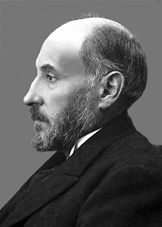
The Nobel Prize in Physiology or Medicine 1906 Camillo Golgi, Santiago Ramón y Cajal

The Nobel Prize in Physiology or Medicine 1906 was awarded jointly to Camillo Golgi and Santiago Ramón y Cajal "in recognition of their work on the structure of the nervous system"

                                                                Santiago Ramón y Cajal
                               http://nobelprize.org/nobel_prizes/medicine/laureates/1906/cajal.html
                                                                         Camillo Golgi
                            http://nobelprize.org/nobel_prizes/medicine/laureates/1906/golgi.html

Exposing the Forest

The 1906 Nobel Prize in Physiology or Medicine was awarded to Camillo Golgi and Santiago Ramón y Cajal for revealing the inner beauty of the nervous system. By developing methods that could colour and highlight its key components, Golgi and Cajal allowed the anatomy of the nervous system to be observed and documented in precise detail.
When Camillo Golgi began studying the grey matter of the brain, existing techniques used to stain cells marked almost all parts of this dense tissue simultaneously, revealing no information about its finer details. Searching for a better staining method, Golgi experimented by candlelight in a hospital kitchen that he converted into a laboratory. There, he discovered a way of impregnating nervous tissue with a silver solution, which stained only a small number of nerve cells in black. This made it possible to view neurons in their entirety, from which Golgi could see the complex outlines and details of numerous branches with remarkable clarity.
Golgi's silver staining method for nerves went unappreciated, until Ramón y Cajal enhanced its resolution and used this technique to investigate brains from young animals. Armed with a microscope, pen and paper, and endowed with artistic talent, Cajal created thousands of beautiful drawings depicting the intricate details of nervous tissue – which to him resembled a forest of outstretched trees. From his observations, Cajal developed the theory that the brain and spinal cord are made up of countless numbers of individual functional units, which his colleague Wilhelm von Waldeyer-Hartz later called neurons. According to this neuron doctrine, these individual cells have their own unique set of nerve processes, and the contents of each cell do not come into direct contact with that of other cells. Each neuron conveys its information to other cells through nerve fibres that extend across the space and barriers that separate them.
However, at the time the Nobel Prize was awarded, Golgi did not support the neuron theory for which Cajal had provided so much evidence – Golgi still interpreted the observations to indicate that the nervous system existed as a seamless continuous network, which forms an entirely distinct anatomical entity. Despite their difference in opinion, Golgi and Cajal became the first scientists to ever share a Nobel Prize.
By Joachim Pietzsch, for Nobelprize.org

Nobel Prize - Neuroscience

The Nobel Prize in Physiology or Medicine (Swedish: Nobelpriset i fysiologi eller medicin) administered by the Nobel Foundation, is awarded once a year for outstanding contributions in the medical field. So From the following website you can actually see people who have received noble prize from the field of neuroscience.

http://faculty.washington.edu/chudler/nobel.html

MRI Scans Reveal Brain Changes in People at Genetic Risk for Alzheimer's

 Identifying high-risk populations is an important component of disease prevention strategies. One approach for identifying at-risk populations for Alzheimer's disease (AD) is examining neuroimaging parameters that differ between patients, including functional connections known to be disrupted within the default-mode network. We have previously shown these same disruptions in cognitively normal elderly who have amyloid-β (Aβ) plaques [detected using Pittsburgh Compound B (PIB) PET imaging], suggesting neuronal toxicity of plaques. Here we sought to determine if pathological effects of apolipoprotein E {varepsilon}4 (APOE4) genotype could be seen independent of Aβ plaque toxicity by examining resting state fMRI functional connectivity (fcMRI) in participants without preclinical fibrillar amyloid deposition (PIB–). Cognitively normal participants enrolled in longitudinal studies (n = 100, mean age = 62) who were PIB– were categorized into those with and without an APOE4 allele and studied using fcMRI. APOE4 allele carriers (E4+) differed significantly from E4– in functional connectivity of the precuneus to several regions previously defined as having abnormal connectivity in a group of AD participants. These effects were observed before any manifestations of cognitive changes and in the absence of brain fibrillar Aβ plaque deposition, suggesting that early manifestations of a genetic effect can be detected using fcMRI and that these changes may antedate the pathological effects of fibrillar amyloid plaque toxicity.


http://www.sciencedaily.com/releases/2010/12/101216122127.htm

Wednesday, December 29, 2010

Functional imaging of hippocampal place cells at cellular resolution during virtual navigation

Spatial navigation is often used as a behavioral task in studies of the neuronal circuits that underlie cognition, learning and memory in rodents. The combination of in vivo microscopy with genetically encoded indicators has provided an important new tool for studying neuronal circuits, but has been technically difficult to apply during navigation. Here we describe methods for imaging the activity of neurons in the CA1 region of the hippocampus with subcellular resolution in behaving mice. Neurons that expressed the genetically encoded calcium indicator GCaMP3 were imaged through a chronic hippocampal window. Head-restrained mice performed spatial behaviors in a setup combining a virtual reality system and a custom-built two-photon microscope. We optically identified populations of place cells and determined the correlation between the location of their place fields in the virtual environment and their anatomical location in the local circuit. The combination of virtual reality and high-resolution functional imaging should allow a new generation of studies to investigate neuronal circuit dynamics during behavior.

http://www.nature.com/neuro/journal/v13/n11/abs/nn.2648.html

Neuroscience As a core

Neuroscience Core Concepts offer fundamental principles that one should know about the brain and nervous system, the most complex living structure known in the universe. They are a practical resource about:
  • How your brain works and how it is formed.
  • How it guides you through the changes in life.
  • Why it is important to increase understanding of the brain.
   we do consciously or unconsciously, our Brain does it very consciously for our safety. Each action is an action potential of a neuron. So this blog is completely dedicated to concepts in neuroscience.

              From behavior ---------------------------------- Cell---------------------------------Gene

Your suggestions ----- quotes------ messages--------comments ---------- are most welcome.

your's
Neurokish